-
 Hydroethidine™ (Dihydroethidium bromide)Catalog Number 17084
Reduced ethidium bromide. A vital stain. Enters and stains living cells without cellular trauma.
-
 Osmium ammine-BCatalog Number 21033
Stable DNA stain.
-
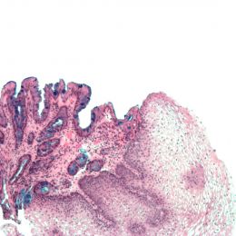
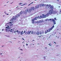
 Toluidine blue O, C.I. 52040, certifiedCatalog Number 01234
A metachromatic, cationic thiazine dye that is widely used in in vitro biological applications. It has also been used in techniques for DNAase detection.
λmax 626nm
-
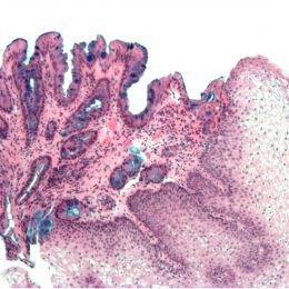
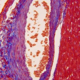
 Toluidine blue O, C.I. 52040, purifiedCatalog Number 15931
Useful for staining RNA, oligodeoxynucleotides, proteins and glycosaminoglycans.
λ max 626nm.
Assay >60% anhydrous dye.
-
 Indocyanine greenCatalog Number 08263
Dye for hemodynamic studies. Invest.
Cardiogreen; Fox green λ max 775nm
-
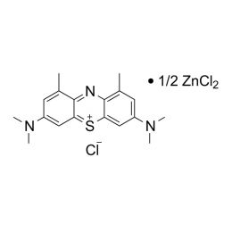 1,9-dimethyl methylene blue zinc chloride double salt (DMMB)Catalog Number 03610
(Taylor’s blue; 3,7’-Bis(dimethylamino)-1,9-dimethyldiphenothiazin-5-ium chloride) λ max 649nm
Used in dye binding assays for glycosaminoglycans.
-
 SaffronCatalog Number 25007
Saffron is used in a variety of histological staining methods including the Hematoxylin, Phloxine and Saffron (HPS) staining technique.
-
 Terry's Polychrome Methylene Blue 2% AqueousCatalog Number 09978
A STAT staining method for unfixed and fixed tissue. Stains nuclei more strongly than cell cytoplasm.Excellent dye for nuclear and nucleolar details. Can be used to demonstrate erythrocyte alterations/inclusions and some erythrocyte parasites, as well as to visualize reticulocytes.
-
 Ruthenium hexammine trichlorideCatalog Number 17253
Electron microscopy stain.
-
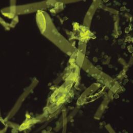
 Uvitex 2BCatalog Number 19517
Uvitex 2B fluorescent dye will bind to the chitin in fungal walls and is highly selective for fungi and algae in tissue sections. It has a slower fade rate than more commonly used fungal fluorescent dyes.
-
 Thiazole orangeCatalog Number 19352
Fluorescent dye for reticulocyte analysis. Also useful for Plasmodium species analysis.
λ max: 512nm
-
 Szechrome NASCatalog Number 08762Inquire for availability.Phone: +(49) 6201-845200Email: [email protected]
Suitable for the determination of the nitrate content of natural waters, industrial waste effluents, soil, plant and meat extracts, tinned goods, biological fluids (sputum, urine), chemicals, fertilizers and drugs. NAS has been used in place of the phenoldisulfonic acid, brucine and chromotropic acid methods used previously.
-
 Fungi-Fluor® Kit for Fungal Detection (for U.S. & outside of Europe orders)Catalog Number 17442Inquire for availability.Phone: +(49) 6201-845200Email: [email protected]
The Fungi-Fluor® Kit for Fungal Detection offers a quick fluorescent stain/counterstain procedure for various fungal organisms. The kit can be used to screen a variety of specimen types, such as sputum and skin scrapings, for fungal detection.
-
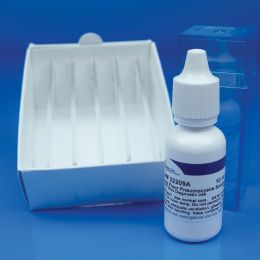 Fungi-Fluor® Pneumocystis Kit (for U.S. & outside of Europe orders)Catalog Number 22363Inquire for availability.Phone: +(49) 6201-845200Email: [email protected]
Pneumocystis jiroveci (previously named P. carinii) is an organism that can cause Pneumocystis carinii pneumonia (PCP) in immunocompromised patients. PCP, long associated with morbidity and mortality with HIV-infected (AIDS) patients, is now increasing in prevalence among the non-HIV infected immunosuppressed population. Early detection allows the introduction of appropriate treatment and may improve chances of patient survival. The Fungi Fluor® Pneumocystis kit offers a fast, fluorescent staining procedure for Pneumocystis carinii in bronchial specimens.
Product is for research use only in Europe.
-
 Gomori's Trichrome Stain KitCatalog Number 24205
Gomori’s One Step Trichrome refers to the multiple stain reaction of this reagent only. However, adequate staining of all cell components requires several other solutions to obtain balance in the tissue. The mordant with Bouin’s Fixative is used to drop the pH and for protein interaction in the section. While this mechanism is not clear the stain works best with this step included.
-
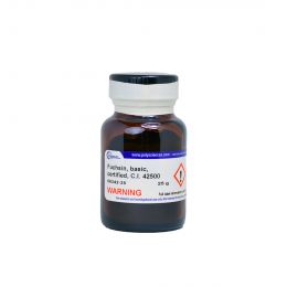 Fuchsin, basic, certified, C.I. 42500Catalog Number 06342
For staining bacilli in tissue. Coupling agent for ultrastructural localization of esterases.
(Pararosaniline hydrochloride; Basic red 9) λ max 542-548nm
-
 Safranin O, C.I. 50240, certifiedCatalog Number 02782
A general biological stain. Used as a nuclear stain for histological studies. Also used as a cationic lipophilic probe and in the detection of glycosaminoglycans and proteoglycans.
(Basic red 2; 3,7-Diamino-2,8-dimethyl-5-phenylphenazinium chloride) λ max 530nm.
-
 Biebrich scarlet, C.I. 26905Catalog Number 03336
Widely used as a counterstain. Also, useful plasma stain.
(acid red 66; Ponceau B) λ max 505nm
-
 Aniline blue, C.I. 42755, Certified, Water-solubleCatalog Number 02570
Used as contrast stain in histology and cytology and as a pH indicator (pH 10.0-13.0). Used with acid fuchsin as Mallory’s connective tissue stain. Also used to visualize chromosomes and cellulose wall implants. (Acid blue 22).
-
 Light green SF yellowish, C.I. 42095, certifiedCatalog Number 02753
Used as a dye and as a biological stain. Certified for use as a counterstain in cytology. An important contrast stain for plasma as a critical component of Papanicolaou (PAP) stains. Stains collagen fibers when substituted for aniline blue in Masson’s trichrome.
λ max 630(422)nm
-
 Chlorazole black E, C.I. 30235, certifiedCatalog Number 02730
A valuable stain in general histology and cytology. Gives sharp, clear-cut pictures of both nuclei and cytoplasmic structures. Also useful for differentiation of fungi.
-
 Tetrachrome stain, certified (MacNeal)Catalog Number 02783
Blood stain similar to Wright’s stain. Also useful for staining bone sections.
-
Nuclear fast red, C.I. 60760Catalog Number 09773
Nuclear fast red staining is a simple method of nuclear chromatin staining and is mainly used for high-contrast counterstaining for histological applications.
-
 Alcian Blue 8GX, C.I. 74240Catalog Number 19175
Used primarily for demonstrating acid mucopolysaccarides with Scott's method and Mowry's staining methods. Used in electrophoresis for detecting glycoproteins.
-
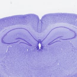 Cresyl violet acetate, certifiedCatalog Number 21063
(9-Amino-5-imino-5H-benzo[a]phenoxazine acetate salt) Cresyl Violet Acetate is a certified solid dye used for the preparation of a staining solution to demonstrate the presence of Nissl substances in neurons and cell nuclei.
-
 Sudan Black B, C.I. 26150, CertifiedCatalog Number 25008
Sudan Black B is useful for staining neutral triglycerides and lipids on frozen sections and some lipoproteins on paraffin sections.
-
 Rose Bengal, C.I. 45440, CertifiedCatalog Number 25005Inquire for availability.Phone: +(49) 6201-845200Email: [email protected]
Rose Bengal can be used as an alternative to phloxine B in Kreyberg's stain for keratin and mucus. Useful for detecting bacteria in soil.
-
Orange G, C.I. 16230, certifiedCatalog Number 00968
Mallory’s stain for collagen in connective tissue. Also useful as a stain for granules, elastic fibers, mast cells, and pollen tubes. A general histology and cytology counterstain.
Acid orange 10; Xylene fast orange G; 7-Hydroxy-8-phenylazo-1,3-naphthalenedisulfonic acid λmax 475nm.
-
 Eosin Y, C.I. 45380, certifiedCatalog Number 02740
Cytoplasmic counterstain. Component of Wright stain and Tetrachrome stain.
(Eosin yellowish; Acid red 87; 2’,4’,5’,7’-Tetrabromo-fluorescein, disodium salt)
λmax: 517nm
-
Congo red, C.I. 22120, certified (Direct red 28)Catalog Number 02736
Anionic metachromatic dye. Good contrast stain or counterstain. Specific stain for amyloids in pathology. Also, a pH indicator; transition interval: pH 3.0 (blue) to 5.0 (red).
λ max: 497nm
-
 Alcian Blue/PAS KitCatalog Number 25086
Demonstrates neutral and acidic mucosubstances on tissue at pH 2.5 imparts a blue color to the acidic mucins and other carboxylated or weakly sulphated acid mucosubstances. Periodic Acid Schiff (PAS) reaction is then used to stain basement membranes, glycogen and neutral mucosubstances pink to red. Mixtures of neutral and acidic mucosubstances will appear purple due to positive reactions with both Alcian Blue and PAS.
-
 Luxol Fast Blue, Ready-to-UseCatalog Number 24611
Used to stain myelin and phospholipids.
-
 Neat Stain Hematology Stain KitCatalog Number 25034
Three-step procedure for differentiation of morphological cell types in peripheral blood smears. Staining characteristics are similar to the traditional Wright’s and Wright-Giemsa stains.
-
 Villanueva Osteochrome Bone StainCatalog Number 16280
Useful for staining fresh, fixed, unembedded or plastic-embedded sections of bone. Villanueva Osteochrome Bone Stain gives uniform and reproducible results. Useful in studying biopsy or postmortem tissue.
-
 Verhoeff Van Gieson Elastin Stain KitCatalog Number 25089
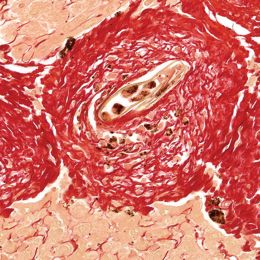
This stain is useful in demonstrating atrophy of elastic tissue in cases of emphysema, thinning and loss of elastic fibers in arteriosclerosis and other vascular diseases. With increasing age, changes such as splitting and fragmentation occur, these changes are most obvious in the skin which becomes wrinkled and rather loose-fitting.
-
Gram Stain Set (Stabilized)Catalog Number 25036
For staining bacteria from cultures or specimens by the differential Gram stain method. Kit performs 150 tests.
-
 Amyloid Stain Kit (Congo Red)Catalog Number 24614
Complete kit to aid in the staining of tissue for Amyloidosis. Amyloid Stain Kit used in the detection of amyloid FFPE as well as frozen tissue sections cut at 10 microns. The amyloid stains red and the nuclei stains blue. Control tissue is Alzheimer’s or other known amyloidosis.
-
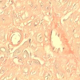
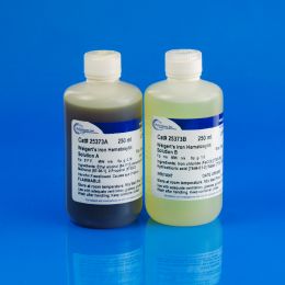 Weigert’s Hematoxylin Kit (Solution A & B)Catalog Number 25373
A nuclear staining solution containing, hematoxylin, ferric chloride and hydrochloric acid used in many non-routine techniques (special stains) because, it resists decolorization in acidic staining solutions. Ferric Chloride is a strong oxidizer, so it serves both as a mordant and oxidizer for Weigert's Hematoxylin.
-
 Fungi-Fluor® Kit for Fungal Detection (for European Orders only)Catalog Number 17442E
The Fungi-Fluor® Kit for Fungal Detection offers a quick fluorescent stain/counterstain procedure for various fungal organisms. The kit can be used to screen a variety of specimen types, such as sputum and skin scrapings, for fungal detection.
-
 Fungi-Fluor® Pneumocystis Kit (for European Orders only)Catalog Number 22363E
Pneumocystis jiroveci (previously named P. carinii) is an organism that can cause Pneumocystis carinii pneumonia (PCP) in immunocompromised patients. PCP, long associated with morbidity and mortality with HIV-infected (AIDS) patients, is now increasing in prevalence among the non-HIV infected immunosuppressed population. Early detection allows the introduction of appropriate treatment and may improve chances of patient survival. The Fungi Fluor® Pneumocystis kit offers a fast, fluorescent staining procedure for Pneumocystis carinii in bronchial specimens.
-
 Nuclear Fast Red, 1% SolutionCatalog Number 24199C
 -
Phosphotungstic Phosphomolybdic AcidCatalog Number 25088D
-
 N-(ɣ-L-Glutamyl)-4-methoxy-2-naphthylamineCatalog Number 02410
Used as a substrate for the histochemical demonstration of γ-glutamyl transpeptidase activity.
-
 Rhodamine 6G, C.I. 45160Catalog Number 25004Inquire for availability.Phone: +(49) 6201-845200Email: [email protected]
Used as a tracer dye within water to determine the rate and direction of flow and transport.
-
 Acid Fuchsin, C.I. 42685, certifiedCatalog Number 24991
Acid Fuchsin is one of the dyes used in the Masson's Trichrome Staining technique. This method is commonly used to stain tissue sections in the Histology and Cytology Laboratory in order to distinguish muscle from collagen. The muscle stains red with the acid fuchsin and the collagen is stained green or blue with light green SF yellowish or methyl blue.
-
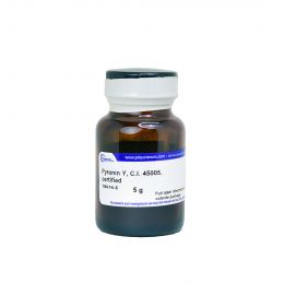 Pyronin Y, C.I. 45005, certifiedCatalog Number 18614
Used in combination with methyl green for the selective and differential staining of nucleic acids. The pyronin Y stains RNA red, while the methyl green stains DNA green. The combined methyl green-pyronin Y stain is a useful histochemical reagent. Pyronin Y can also be used as a tracking dye for polyacrylamide gel electrophoresis.
λmax 552nm
-
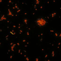 Acridine orange, C.I. 46005 (min. 95%)Catalog Number 04539
DNA intercalating dye. A grade of acridine orange of exceptionally high purity, suitable for quantitative work. Free of inorganic salts. A specific stain for RNA, used as a 2% solution containing 1% lanthanum acetate in 15% acetic acid.
-
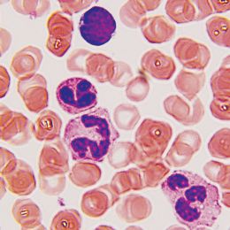
 Wright stain, certifiedCatalog Number 02785
Useful for staining blood films and malarial parasites in blood films.
-
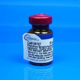 Filipin (from Streptomyces filipinensis)Catalog Number 08707
Polyene antibiotic fluorochrome for cholesterol determination. Used as an antifungal agent.
-
Oil Red O, C.I. 26125, certifiedCatalog Number 06317
A post-electrophoresis stain for lipoproteins on cellulose acetate plates.
(Solvent red 27; Sudan red 5B; Ceres red 5B)
-
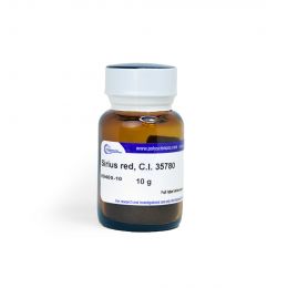 Sirius red, C.I. 35780Catalog Number 09400
Cardiac muscle stain.
(Direct red 80)
-
 Fast green FCF, C.I. 42053, certifiedCatalog Number 02745
(Food green 3). Sensitive stain for proteins in polyacrylamide gels. Especially suitable in isoelectric focusing. Also suitable for use as a cytological counterstain, and mammalian tissue stain for muscle and collagen.
λ max 622-626nm.
-
 Coomassie® Blue G250, C.I. 42655Catalog Number 03707
Protein stain for SDS gels. Used in dye binding techniques for protein quantification.
(Coomassie blue® G250; Brilliant blue G250; Acid blue 90)
λmax 610nm
-
 Coomassie® blue R250, C.I. 42660Catalog Number 00352
Rapid acting and sensitive dye for SDS gels.
(Coomassie blue® R250; Brilliant blue R250; Acid blue 83)
λmax 588nm
-
 Hematoxylin, C.I. 75290, certified (Natural black 1)Catalog Number 02749
Nuclear protein stain and glycogen stain. Also a general tissue stain for human, animal and VIR histology.
-
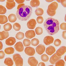 StainRITE® Wright Stain SolutionCatalog Number 24986
Wright Stain is a dual purpose stain used for staining blood smears and bone marrow aspirates. Ready-to-use solution makes the differentiation of human blood cells much easier to identify.
-
 Multiple Stain SolutionCatalog Number 08824
Used in Tzanck preparations of herpetic lesions and to differentiate acidophilic and basophilic structures. Stain can be directly applied to frozen sections, epoxy or JB-4® embedded sections and utilized as a general cytoplasmic and nuclear stain.
-
 Gill’s modified OG-6Catalog Number 09782
OG-6 is a cytoplasmic counterstain solution used in sequence with EA (EA 50, EA 65 or EA36) in the Papanicolaou staining method for clinical cytology. Gill’s modified OG-6 is stable in solution and gives predictable high quality staining results not previously possible with other formulations.
-
 Gill’s modified EACatalog Number 09783
Orange-6 and EA are the two cytoplasmic counterstain solutions that are used sequentially in the Papanicolaou staining method for clinical cytology. Gill’s modified OG-6 and EA are stable in solution and give predictable high quality staining results not previously possible with other formulations.
-
 Picrosirius Red Stain KitCatalog Number 24901
Collagen Type I and III Staining
Our Picrosirius Red Stain binds specifically to collagen fibrils of varying diameter that is used to distinguish collagen Type I from Type III. Picrosirius Red Stain will quantify the amount of collagen in a given area of myocardial tissue. (i.e. the collagen area fraction)
-
StainRITE® Giemsa Stain (for May-Grünwald)Catalog Number 25038
Giemsa stain is a classical blood film stain for peripheral blood smears and bone marrow specimens, used to visualize chromosomes, stains fungus histoplasma and identifies mast cells.
To be used in conjunction with our May-Grünwald Stain Solution, Cat. #24981
-
 StainRITE® Wright-Giemsa Stain SolutionCatalog Number 24985
Wright-Giemsa Stain Solution is a dual purpose stain useful for blood films, parasites and bone marrow aspirates. Prepared from certified dyes.
Ready-to-use solution makes the differentiation of human blood cells much easier to identify.
-
 Scott's Bluing ReagentCatalog Number 24605
A gentle formulation of "bluing" reagent for those specimens that may be affected by more harsh "bluing" agents. Our premixed and ready to use Scott's Bluing Reagent provides rapid bluing and crisp nuclear detail obtained with routine Hematoxylin and Eosin stains. For use in both Cytology and Histology.
-
 Masson's Trichrome Stain KitCatalog Number 25088
Used for the detection of collagen fibers in tissues such as skin, heart, etc. on formalin-fixed, paraffin-embedded sections and may be used for frozen sections as well. Collagen fibers stained blue, nuclei stained black and cytoplasm, muscle, erythrocytes stained red.
Available in 100ml and 500ml
-
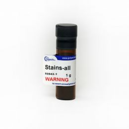 Stains-allCatalog Number 03943
Used to stain DNA, RNA, and protein. Also useful for staining acid polysaccharides.
Results:DNA stain blue
RNA stain bluish-purple
Protein stain red
-
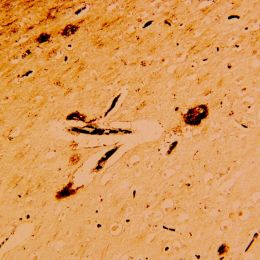 Bielschowsky Stain KitCatalog Number 25994
The Bielschowsky Stain is a very useful method in detecting the twist and clumping of neurons in the brain, otherwise known as, neurofibrillary tangles and senile plaques, which are signs of the Alzheimer’s disease. The Bielschowsky stain, along with other diagnostic test, can assist scientist and researchers in the mission for finding a cure for this disease of memory loss.
-
 Phosphomolybdic acid hydrate, ACS gradeCatalog Number 01021
Electron dense metal stain.
(12-Molybdophosphoric acid)
-
 Oil Red O Stain KitCatalog Number 25962
Oil Red O is excellent for the demonstration of general localization of fats in frozen tissue sections.
-
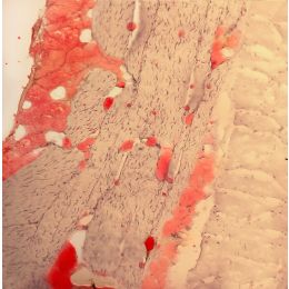
 Prussian Blue Iron Stain Kit (Reaction for Demonstration of Iron)Catalog Number 24199
Prussian Blue or Perls’ reaction is used to demonstrate ferric iron and ferritin. This is not a true staining technique rather, it is a histochemical reaction.
The protein is split off by the hydrochloric acid, allowing the potassium ferrocyanide to combine with the ferric iron. This forms the ferric ferrocyanide or Prussian Blue.
-
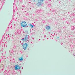
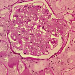 Periodic Acid Schiff's (PAS) Stain KitCatalog Number 24200
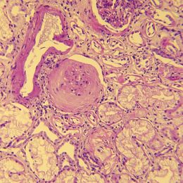
PAS techniques are used to demonstrate polysaccharides, neutral mucosubstances and basement membranes primarily in tissue. The PAS reagent is also called Fuelgen Stain for the demonstration of DNA with a different protocol. Kidney is the most sensitive control. The demonstration of glycogen is best represented by a section of liver with a digestion step used as a negative control in the staining.
Stains, Dyes & Indicators - Histology